


What does the new coronavirus look like and how to take pictures of the virus?
Release time:
2020-10-23
The new crown virus is raging around the world and can be said to be the most terrifying enemy of mankind. So what does this virus look like? Some people say that since it is a coronavirus, it certainly looks like a crown, which is true, but the pictures on the Internet are all mock or conceptual pictures, not the actual appearance or photos of the virus.
So how can you see the real look of the virus or take pictures of the virus? Of course it is a microscope, but ordinary microscopes cannot. The resolution of the microscope is limited, and the magnification does not exceed 2000 times. The maximum can only see objects the size of cells. It is difficult to see the organelles, and the virus is much smaller than many organelles.
Observing objects smaller than 200nm with a microscope is completely blurred or invisible. We know that light travels in a straight line. In fact, for the microscopic world, light does not always travel along a straight line. It will reflect the volatility and diffract. The wavelength of visible light is 380-760nm, and the ultraviolet light will be lower, but for the scale below 100nm, it is related to the wavelength of light. Quite or even shorter, light cannot be clearly imaged by reflection or transmission.
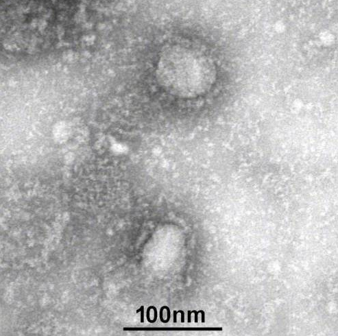
Virus electron micrograph
With the development of science, scientists have developed electron microscopes. Unlike ordinary microscopes, electrons cannot be magnified and imaged through a lens. Instead, electric or magnetic fields are used to change the electron trajectory to form a magnification effect. This electric or magnetic field is equivalent to the function of an electronic lens. Electron is different from photon, it has quality, so the wavelength is much smaller than light wave, the resolution is greatly improved, electron beam replaces visible light, the resolution reaches 0.1nm, this technology won the Nobel Prize in 1986.
The disadvantage of the electron microscope is that it is a black and white world, because the image it obtains is a "gray image" reflecting the amount of electrons (ie brightness). Unlike visible light, which has different colors, we can also easily give specific biological tissues. The ingredients are dyed or fluorescently labeled. The post-coloring of the picture can make the electron microscope photos appear in false colors, but the photos with little grayscale difference can also be distinguished by Hannan.
The development of technology is achieved by countless scientific workers' hard work and accumulation. The coloring technology of electron microscopy photos is also constantly developing. I believe that in the near future we will be able to see high-definition color photos of viruses, even DNA molecules or single proteins. photo. Desheng is a manufacturer of sampling tubes, virus transport medium, and biological buffers related to virus detection, and colleagues are welcome to exchange and learn from each other.
Contact details
Contact number
Address: C8, Guanggu United Science and Technology City, Ezhou City, Hubei Province
Fax:0711-3704 589
Follow us
